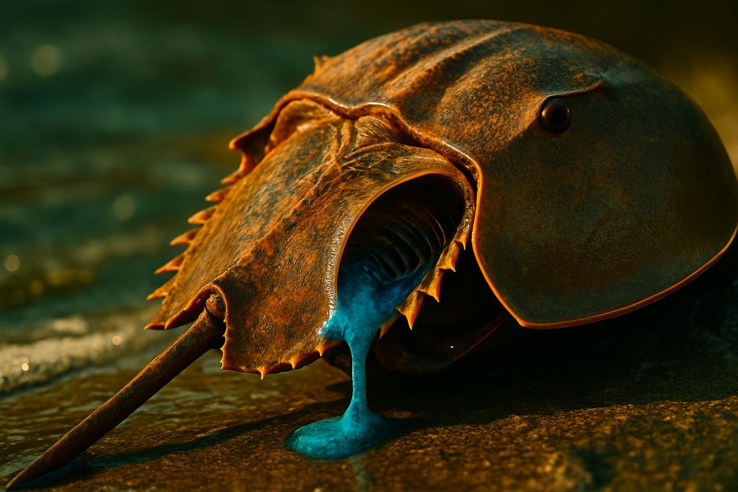
A single drop of bacterial toxin dissolved into 20 Olympic swimming pools — that’s the detection threshold horseshoe crab blood has been running on for 450 million years. Every injectable drug cleared for human use passes through a test built around this same cascade.
Horseshoe crab blood contains specialized immune cells called amebocytes that trigger a rapid clotting cascade when they encounter bacterial endotoxin, and that biological mechanism became the worldwide standard test for detecting contamination in injectable drugs, vaccines, and medical devices. The cascade doesn’t involve antibodies or immune memory. It’s a purely chemical chain reaction — fast, ancient, and extraordinarily sensitive.
The moment lipopolysaccharide (LPS), the endotoxin shed by Gram-negative bacteria, enters a horseshoe crab’s bloodstream, amebocytes rupture and release clotting proteins. Those proteins form an insoluble gel around the invader within minutes, trapping and isolating it. The crab’s blood turns the pathogen into a walled-off problem rather than a spreading one.
Quick Facts
– Horseshoe crabs have circulated on Earth for roughly 450 million years
– Detection sensitivity reaches one part per trillion — confirmed as low as 45 minutes per turnaround
– The clotting cascade runs through factor C, factor B, proclotting enzyme, and coagulogen
– Fungi trigger a separate parallel pathway via factor G, activated by beta-(1,3) glucan
– The test is called the Limulus Amebocyte Lysate (LAL) assay
How the Horseshoe Crab Clotting Cascade Actually Works
LPS activates factor C, the first serine protease zymogen in the chain. Factor C then activates factor B, which activates the proclotting enzyme. That enzyme converts coagulogen into coagulin — the insoluble gel. Three serine protease zymogens drive the entire cascade, each one amplifying the signal downstream. The gel doesn’t just wall off the bacterium; it also concentrates antimicrobial substances around it.
Fungi trigger a separate but parallel pathway. Beta-(1,3) glucan, a component of fungal cell walls, activates factor G instead of factor C, feeding into the same downstream clotting machinery. One cascade, two entry points.
How the LAL Test Was Developed and What It Tests Today
Frederick Bang and Jack Levin observed in the 1960s and early 1970s that horseshoe crab blood clotted in the presence of bacterial contamination. By 1971, the connection to LPS from E. coli had been documented. The Limulus Amebocyte Lysate test — named for the crab’s genus, Limulus polyphemus — grew from those observations.
Today the LAL test screens injectable drugs, vaccines, intravenous fluids, and implantable medical devices for endotoxin contamination. Sensitivity reaches one part per trillion — roughly one drop in 20 Olympic swimming pools — with results readable in as little as 45 minutes, though some protocols allow a few hours.
Closing
The LAL test remains the global standard not because nothing else was ever attempted, but because the crab’s cascade proved exceptionally difficult to match on sensitivity and speed. A lineage that survived five mass extinctions, still running the same molecular alarm system it refined hundreds of millions of years ago, now sits at the center of modern pharmaceutical safety.
Some mechanisms don’t get replaced. They just get borrowed.
Frequently Asked Questions
What makes horseshoe crab blood blue?
Horseshoe crab blood contains hemocyanin, a copper-based oxygen carrier, instead of the iron-based hemoglobin found in human blood.
What bacterial toxin does the LAL test detect?
It primarily detects lipopolysaccharide (LPS), the endotoxin present in the outer membrane of Gram-negative bacteria.
How sensitive is the LAL test?
One verified source places detection as low as one part per trillion, with results confirmed in as little as 45 minutes.
When was the LAL test developed?
Research by Frederick Bang and Jack Levin in the 1960s and early 1970s established the foundation; the clotting response to E. coli was documented by 1971.